Dental Fluorosis: White Marks From Too Much Fluoride

White spots, streaks, or faint chalky marks on the teeth that were always there, going back as far as you can remember, are often the result of dental fluorosis. Most people who have it have no idea what it is called or what caused it, and they sometimes worry it is a sign of decay or a problem with their tooth enamel. Dental fluorosis is neither of those things. It is a cosmetic condition that developed during childhood, is entirely benign in its milder forms, and has straightforward treatment options when the appearance bothers a patient.
At Willow Family Dentistry in Wylie, TX, Dr. Esther B. Jeong discusses dental fluorosis with patients who notice it on their own teeth or on their children's teeth and can advise on when cosmetic treatment makes sense.
What Is Dental Fluorosis?
Dental fluorosis is a cosmetic change in tooth enamel caused by excess fluoride intake during the years when permanent teeth were developing. It produces white spots, streaks, or chalky marks that were present when the teeth erupted and do not indicate decay or structural weakness.
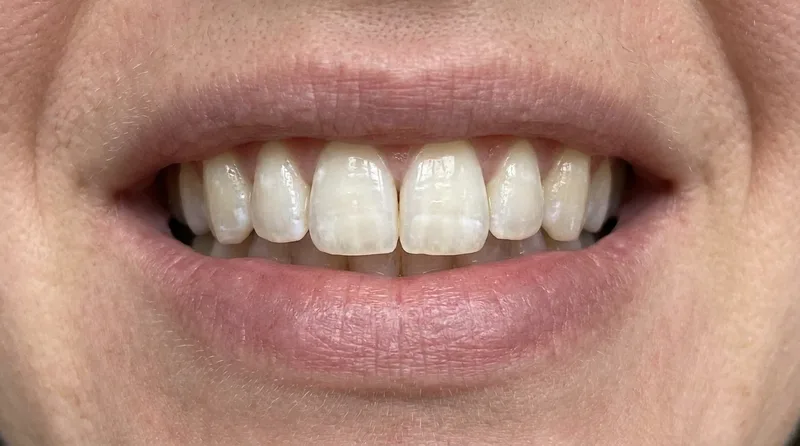
Dental fluorosis is a change in the appearance of tooth enamel caused by excessive fluoride intake during the years when the permanent teeth were developing, typically from birth through about age eight. The fluoride disrupts the process of enamel mineralization during tooth formation, producing enamel that is less translucent than normal and that can range from faint white flecks to more pronounced white streaks to, in severe cases, pitting and brown staining.
Fluorosis only affects teeth that were forming at the time of excess fluoride exposure. It does not develop in permanent teeth after they have already erupted, and it cannot develop in adult teeth regardless of fluoride intake. The condition is cosmetic rather than pathological: fluorosed teeth are not structurally weaker, not more cavity-prone, and do not require treatment on medical grounds. The only reason to treat dental fluorosis is the patient's preference regarding appearance.
According to the National Institute of Dental and Craniofacial Research, most dental fluorosis in the United States is mild and does not affect dental health or function, with only a small proportion of cases producing the more visible pitting or brown discoloration that patients typically find cosmetically significant.
What Causes Dental Fluorosis?
Dental fluorosis is caused by excess fluoride ingestion during childhood, typically before age eight when permanent teeth are mineralizing. The most common sources are swallowing fluoride toothpaste, naturally high fluoride in well water, and fluoride supplementation exceeding the level appropriate for local water content.
Dental fluorosis is caused by ingesting too much fluoride during the developmental years, particularly before age eight when permanent teeth are mineralizing beneath the gum. The most common sources of excess fluoride in children are swallowing fluoride toothpaste rather than spitting it out, living in an area with naturally high fluoride in the water supply, and receiving more fluoride supplementation than appropriate for the local water fluoride level.
In the United States, community water fluoridation is adjusted to a level that supports dental health without causing fluorosis at meaningful rates. According to the Centers for Disease Control and Prevention, community water fluoridation is one of the ten great public health achievements of the twentieth century, and the current recommended level of 0.7 parts per million is calibrated to minimize fluorosis risk while preserving cavity prevention benefit. However, children who live in areas with naturally higher fluoride concentrations in well water, or who swallow fluoride toothpaste consistently during brushing, can exceed the threshold that produces mild fluorosis.
Research has found that dental fluorosis prevalence in the United States increased significantly between the 1980s and 2000s, with some studies estimating that approximately 23% of Americans aged 6 to 49 have some degree of dental fluorosis, according to that epidemiological data. The majority of those cases are very mild, producing only faint white spots that most patients never notice themselves.
What Does Dental Fluorosis Look Like?
Dental fluorosis presents across a severity spectrum that ranges from barely perceptible to moderately significant. The appearance depends on when and how much excess fluoride was ingested during development, and on which specific teeth were forming during that period.
The spectrum from least to most significant:
- Questionable or very mild: A few isolated white flecks or specks on otherwise normally appearing teeth. Often noticed only by a dentist during examination. Many patients with this degree of fluorosis never know they have it.
- Mild: White, opaque areas covering less than 25% of the tooth surface. The enamel appears slightly chalky or cloudy in certain lighting. Visible if someone looks closely, but not typically the first thing people notice in a smile.
- Moderate: White or chalky areas covering more than 25% of the tooth surface, sometimes with a yellowish or brownish tint that develops as the porous fluorosed enamel absorbs staining over time. Visible in natural light and often the reason patients bring it up at a dental appointment.
- Severe: Widespread surface disruption including pitting, rough texture, and brown or black staining. This level of fluorosis is uncommon in the United States under current water fluoridation standards and typically occurs only with very high fluoride exposure.
Fluorosis appears in a characteristic pattern on the teeth: it is bilateral and roughly symmetrical, appearing on the same teeth on both sides of the mouth, because both sides were forming at the same time when the excess fluoride exposure occurred. This symmetry helps distinguish fluorosis from other causes of white spots such as early decay lesions, which typically appear at random locations on individual teeth.

How Is Dental Fluorosis Different From Early Decay?
The most common concern patients raise when they notice white spots on their teeth is whether they are looking at cavities. Dental fluorosis and early decay lesions can both appear as white spots, but they have distinguishing characteristics a dentist can use to tell them apart.
Fluorosis spots are developmental, meaning they were present when the tooth erupted and have been there ever since. They appear in a symmetrical distribution across corresponding teeth. The surface texture is typically smooth, not chalky or soft. Early decay lesions, by contrast, develop on erupted teeth, tend to appear at areas of plaque accumulation like the gumline and between teeth, feel slightly soft or rough to a dental probe, and are not bilateral.
Our article on enamel erosion covers how acid-driven surface changes differ from the developmental change of fluorosis, and our article on teeth sensitive to sweets covers the sensitivity patterns that accompany active decay versus the typically non-sensitive surface of fluorosed enamel.
Can Dental Fluorosis Be Treated?
Dental fluorosis can be improved cosmetically through several approaches depending on the severity and extent of the discoloration. None of these treatments address an underlying health problem, because fluorosis is not a health problem. They address appearance only, and the decision to treat is entirely the patient's.
Treatment options in roughly ascending order of intervention:
- Microabrasion: A mildly abrasive paste applied to the affected enamel surface to remove the outermost fluorosed layer. Effective for mild to moderate fluorosis with surface-level discoloration. Quick, in-office procedure requiring no anesthesia.
- Tooth whitening: Professional whitening can improve the overall appearance of fluorosed teeth and reduce the contrast between the white spots and the surrounding enamel. It does not remove the fluorosis itself but can make the affected areas less conspicuous against a uniformly brighter background.
- Composite bonding: Tooth-colored resin applied to cover the fluorosed areas and restore a uniform surface appearance. Suitable for moderate fluorosis where microabrasion alone is insufficient. Requires no preparation of healthy tooth structure.
- Veneers: Porcelain or composite veneers cover the tooth surface completely and are the most reliable option for moderate to severe fluorosis affecting most of the visible surface. Appropriate when other approaches would not achieve adequate coverage.
For children who are still developing their permanent teeth, the priority is preventing further fluorosis rather than treating what is already present. Using a pea-sized amount of fluoride toothpaste and teaching children to spit rather than swallow is the most effective preventive measure. Our article on plaque and tartar covers the role of fluoride in cavity prevention alongside the proper use that avoids excess ingestion.
When Should You See a Dentist About Fluorosis?
Dental fluorosis does not require a dental visit on medical grounds. See a dentist if the appearance is bothersome enough to consider treatment, if you are unsure whether the spots are fluorosis or early decay, or if your child has noticeable white marks you want evaluated.
Dental fluorosis does not require a dental visit on medical grounds since it is not a health condition. A dental visit is appropriate if the appearance of the fluorosed areas is bothersome enough to consider treatment, if you are unsure whether what you are seeing is fluorosis or something else, or if your child has noticeable white spots that you want evaluated to confirm they are not early cavities.
At Willow Family Dentistry, Dr. Jeong can confirm the diagnosis visually, explain where on the severity spectrum the case falls, and discuss whether any cosmetic intervention is warranted and which approach would be most appropriate for the degree of change present. Willow Family Dentistry provides preventive and cosmetic dental care for families throughout Wylie and the surrounding North Texas communities.
Questions about white marks on your teeth?
Book an evaluation at Willow Family Dentistry in Wylie, TX. Dr. Jeong can confirm whether what you are seeing is dental fluorosis and advise on treatment options if appearance is a concern.
Explore preventive dentistryFurther Reading
Dental fluorosis connects to several other topics related to tooth enamel and surface appearance.
- Are Dental X-Rays Safe? How Often Do You Need Them?
- Acid Reflux and Your Teeth: The GERD-Dental Connection
- Tooth Notch Gumline: Abfraction Explained
Results may vary. Please consult with Dr. Jeong for personalized treatment recommendations.
Dr. Esther B. Jeong, DDS
DDS · Willow Family Dentistry
Wylie family dentist with 15+ years of experience providing gentle, judgment-free dental care.
Frequently Asked Questions
Was this article helpful?
You may also like



Have a dental question?
Schedule a consultation and get personalized answers from Dr. Jeong.
Call us
(972) 881-0715
Hours
Mon – Thu: 9am – 5pm
Fri: By Appointment
Location
1125 W FM 544, Wylie
Emergency? Same-day appointments available.