Impacted Wisdom Teeth Symptoms: Risks and When to Act
Impacted wisdom teeth symptoms are easy to dismiss because they come and go. Your jaw aches for a few days, then stops. The gum behind your last molar swells, then settles down. You taste something foul in the back of your mouth, rinse with mouthwash, and forget about it. The pattern repeats every few weeks or months, each episode slightly worse than the last, until the day the pain doesn't stop or the swelling doesn't go down. The American Association of Oral and Maxillofacial Surgeons estimates that 85% of wisdom teeth eventually need extraction, and impacted teeth that produce intermittent symptoms are signaling that the clock is ticking.
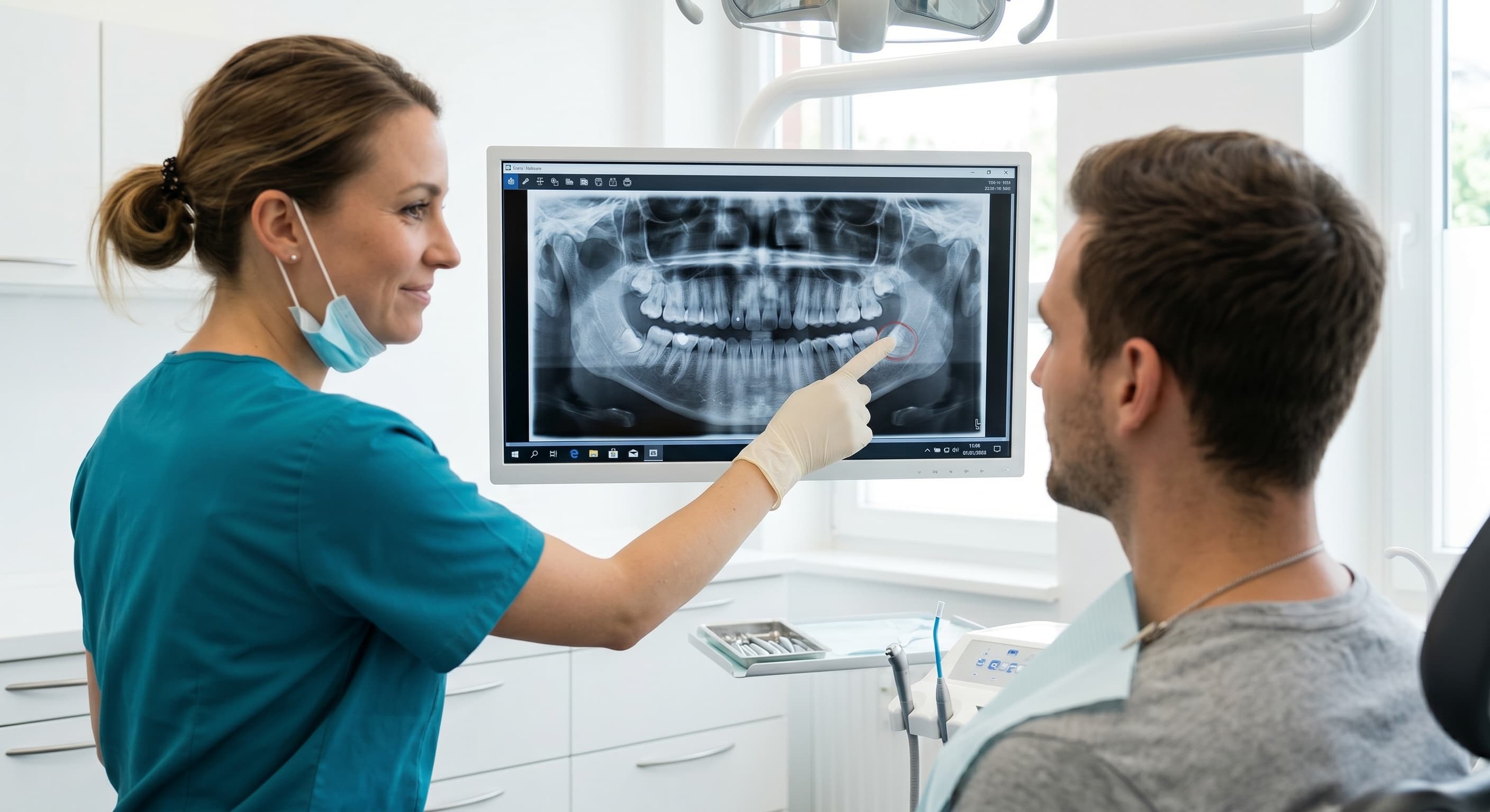
Dr. Esther Jeong at Willow Family Dentistry in Wylie, TX uses iCAT 3D imaging to evaluate impacted wisdom teeth with a level of diagnostic precision that standard X-rays can't match. The 3D scan shows exactly where the tooth sits, how it's angled, whether it's pressing on the adjacent molar, and how close it is to the inferior alveolar nerve. That information determines whether the tooth needs to come out now, can be monitored, or is positioned safely enough to leave alone.
What Are the Symptoms of Impacted Wisdom Teeth?
Impacted wisdom teeth symptoms range from subtle and intermittent to severe and constant. The symptoms depend on the type of impaction (partial vs full, soft tissue vs bony) and whether infection, crowding, or cyst formation has developed. Here are the seven most common symptoms Dr. Jeong evaluates.
1. Pain or Aching at the Back of the Jaw
A dull, persistent ache in the area behind your last molar is the most frequently reported symptom. The pain may radiate to the ear, temple, or along the jawline. It's often worse when chewing, clenching, or opening wide. According to the Mayo Clinic, jaw pain from impaction results from the tooth pressing against bone, the adjacent tooth, or the nerve bundle as it attempts to erupt through tissue it can't get past. The pain is intermittent early on (flaring for days then subsiding) and becomes more constant as the impaction worsens.
2. Swollen, Red, or Tender Gum Behind the Last Molar
When a wisdom tooth is partially erupted, a flap of gum tissue (operculum) covers part of the tooth. Food debris and bacteria collect under this flap in a space that's impossible to clean with brushing or flossing. The trapped bacteria cause localized infection called pericoronitis: the gum becomes swollen, red, tender to touch, and may bleed when you brush near it. Pericoronitis episodes recur every few weeks until the tooth is removed or fully erupts (which impacted teeth can't do).
3. Bad Taste or Smell from the Back of the Mouth
The bacterial pocket around a partially impacted tooth produces a persistent foul taste that no amount of brushing, flossing, or mouthwash fully eliminates. The taste is most noticeable in the morning after overnight bacterial accumulation. If the infection produces pus, you may notice a salty or metallic taste when you press on the gum tissue behind the last molar. According to the ADA, persistent bad taste localized to the wisdom tooth area is a reliable indicator of chronic pericoronitis.
4. Difficulty or Pain When Opening the Mouth
Impaction-related swelling and infection can involve the muscles that control jaw opening (the masseter and medial pterygoid). When these muscles become inflamed, jaw opening becomes restricted and painful, a condition called trismus. You may notice that you can't open wide enough to eat a sandwich or that yawning triggers sharp pain. Trismus associated with wisdom teeth typically indicates significant swelling or infection that has spread beyond the immediate tooth area.
5. Swelling in the Cheek, Jaw, or Neck
Visible facial swelling on the side of the impacted tooth signals that infection has progressed beyond the gum tissue into the surrounding spaces. Mild swelling confined to the gum is early-stage pericoronitis. Swelling that extends to the cheek, angle of the jaw, or neck indicates a spreading infection (cellulitis) that may require antibiotics before extraction can be safely performed. The AAOMS recommends urgent evaluation for any facial swelling associated with wisdom teeth, as dental infections can spread to the airway in rare but serious cases.
6. Crowding or Shifting of Adjacent Teeth
An impacted wisdom tooth angled toward the second molar (mesioangular impaction, the most common type) exerts pressure on the adjacent tooth. Over months or years, this pressure can shift the second molar forward, disrupt the bite, and cause crowding of the front teeth that undermines previous orthodontic work. According to clinical research, patients who notice lower front teeth crowding in their 20s after having straight teeth as teens often have mesioangular impacted wisdom teeth contributing to the shift.
7. Headaches and Referred Pain
Impacted wisdom teeth can trigger headaches through two mechanisms: direct pressure on the trigeminal nerve branches that serve the jaw, face, and temple, and muscle tension from chronic clenching and guarding against the pain. The headaches are typically one-sided (matching the side of the impaction), concentrated at the temple or behind the eye, and worse in the morning if nighttime clenching is involved. Patients often treat these headaches with ibuprofen for months without connecting them to their wisdom teeth.
Recognizing Any of These Symptoms?
Dr. Jeong uses iCAT 3D imaging to see exactly where impacted wisdom teeth sit, how they're affecting adjacent structures, and whether extraction is needed. One scan, complete answers.
Request an Appointment →What Are the Types of Wisdom Tooth Impaction?
The type of impaction determines the symptoms you experience and the surgical complexity of the extraction. Dr. Jeong classifies impaction on the iCAT scan by angulation and depth.
| Impaction Type | Position | Frequency | Risk Level |
|---|---|---|---|
| Mesioangular | Angled forward toward the second molar | Most common (~40%) | High — presses on adjacent tooth, causes crowding |
| Vertical | Upright but trapped beneath gum or bone | Common (~35%) | Moderate — may erupt partially, causing pericoronitis |
| Horizontal | Lying on its side, perpendicular to other teeth | Less common (~15%) | High — can damage second molar root, complex extraction |
| Distoangular | Angled backward toward the jaw ramus | Least common (~10%) | Variable — extraction can be more complex due to angle |
Depth classification adds another dimension: soft tissue impaction (tooth is through the bone but covered by gum), partial bony impaction (tooth is partially covered by bone), and full bony impaction (tooth is completely encased in bone). Full bony impactions often produce no symptoms for years because the tooth is sealed from the oral environment. But they carry the highest risk for cyst formation because the follicular sac surrounding the tooth can fill with fluid and expand within the bone.
What Are the Risks of Leaving Impacted Wisdom Teeth?
The risks of leaving symptomatic impacted wisdom teeth untreated escalate over time. Each risk has a specific mechanism that Dr. Jeong evaluates on the iCAT scan.
Damage to the adjacent second molar. A mesioangular or horizontal impaction pressing against the second molar can cause root resorption (the second molar's root dissolves where the wisdom tooth contacts it), cavity formation on the back surface of the second molar (impossible to treat without removing the wisdom tooth first), and bone loss between the two teeth that compromises the second molar's support. According to the Mayo Clinic, damage to the second molar is one of the strongest indications for wisdom tooth removal even when the wisdom tooth itself isn't painful.
Recurrent pericoronitis. Each episode of infection around a partially impacted tooth risks spreading to the surrounding tissue planes. Mild pericoronitis (gum swelling and tenderness) can progress to severe pericoronitis (facial swelling, trismus, difficulty swallowing, fever) that requires IV antibiotics and urgent surgical drainage. Recurrent episodes three or more times in a year is a clear indication for extraction.
Cyst or tumor formation. The follicular sac surrounding any impacted tooth can develop into a dentigerous cyst that slowly expands and destroys surrounding bone. Cysts are detected on imaging before they cause symptoms, which is why the ADA recommends periodic radiographic monitoring of impacted teeth that are being observed rather than extracted. In rare cases, the cyst lining can undergo malignant transformation (ameloblastoma), making early detection and removal of expanding cysts critical.
Crowding and orthodontic relapse. The forward pressure from mesioangular impacted wisdom teeth contributes to lower anterior crowding over time. Patients who had orthodontic treatment as teens and notice their lower front teeth shifting in their 20s often have this dynamic at work. Removing the impacted teeth doesn't reverse the crowding that's already occurred, but it removes the force that's driving it.
Related: Full procedure walkthrough if extraction is needed. → Wisdom Teeth Removal: What to Expect Before, During, and After
How Does iCAT 3D Imaging Change the Diagnosis?
A standard 2D panoramic X-ray shows impacted wisdom teeth as flat silhouettes. It reveals their general position and angulation but compresses three-dimensional anatomy into a two-dimensional image, which creates blind spots.
The iCAT 3D scan at Willow eliminates those blind spots. Dr. Jeong can measure the exact distance between the wisdom tooth root and the inferior alveolar nerve (the nerve responsible for lower lip and chin sensation) in millimeters. She can see whether the roots wrap around the nerve canal or simply pass near it. She can evaluate bone density around the tooth, identify early cyst formation, and assess the second molar's root for resorption that's invisible on 2D film.
This matters clinically because nerve proximity determines the surgical approach. A tooth with roots 3mm from the nerve is extracted with a standard technique. A tooth with roots intimately associated with the nerve may require a coronectomy (removing the crown and leaving the roots in place to avoid nerve damage) or a modified surgical approach. According to oral surgery research, 3D imaging prior to wisdom tooth extraction reduces the incidence of nerve injury by allowing surgeons to plan the extraction path precisely.
When Should You Act on Impacted Wisdom Teeth Symptoms?
The decision to extract isn't always about current symptoms. It's about the trajectory.
Act now when you have recurrent pericoronitis (three or more episodes), constant rather than intermittent pain, visible facial swelling or trismus, a bad taste that persists despite good hygiene, or imaging that shows damage to the adjacent second molar or cyst formation.
Monitor with regular imaging when the tooth is fully impacted, asymptomatic, and showing no cyst formation or effect on adjacent teeth. Some fully bony impacted wisdom teeth remain harmless for decades. But monitoring means actual periodic imaging (every 1-2 years), not ignoring them and hoping for the best.
Consider proactive extraction when you're in your late teens or early 20s and the roots haven't fully formed yet. Younger patients heal faster, have fewer complications, and the roots are shorter and less curved, making the extraction simpler. The AAOMS recommends evaluation by age 17 so that extraction timing can be optimized for the patient's anatomy and development.
Related: Anxious about the procedure? → Dentist for Anxious Patients in Wylie, TX
Impacted wisdom teeth symptoms are your body's warning system. The intermittent pain, the swelling that comes and goes, the bad taste that won't quit, these aren't random annoyances. They're signals that a tooth is stuck where it shouldn't be, and the longer it stays, the more damage it can do to the bone, the adjacent teeth, and the surrounding tissue. If the symptoms described above sound familiar, schedule an evaluation with Dr. Jeong at Willow Family Dentistry. The iCAT 3D scan shows you exactly what's happening beneath the gum, and the answer, whether it's extraction, monitoring, or nothing, comes from data rather than guesswork.
See What's Happening Beneath the Gum
Dr. Jeong uses iCAT 3D imaging to evaluate impacted wisdom teeth with precision that standard X-rays can't match. One scan reveals position, nerve proximity, and adjacent tooth damage.
Request a Consultation →Jaw pain or swelling behind your last molar?
Call (972) 881-0715 →Dr. Esther B. Jeong, DDS
DDS · Willow Family Dentistry
Wylie family dentist with 15+ years of experience providing gentle, judgment-free dental care.
Frequently Asked Questions
Was this article helpful?


Have a dental question?
Schedule a consultation and get personalized answers from Dr. Jeong.
Call us
(972) 881-0715
Hours
Mon – Thu: 9am – 5pm
Fri: By Appointment
Location
1125 W FM 544, Wylie
Emergency? Same-day appointments available.